
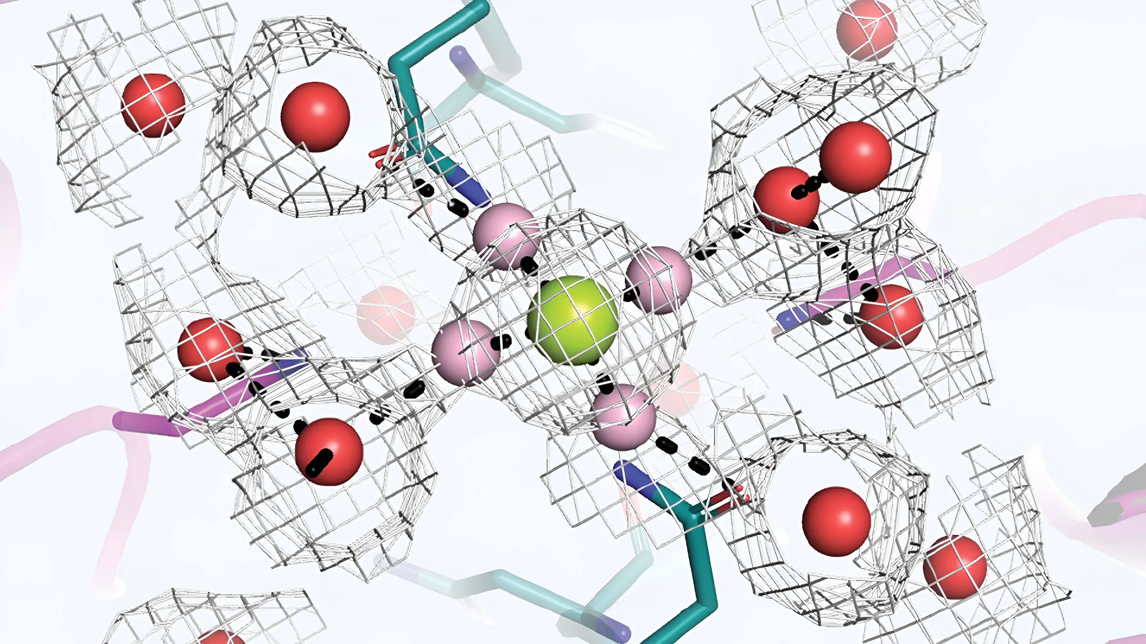
A Cold Spring Harbor Laboratory team has captured, for the first time, the exact molecular process by which a critical brain receptor tells calcium from magnesium — a distinction that sits at the physical foundation of how neurons store memories.
Why Two Nearly Identical Ions Have Opposite Jobs at the Synapse
The receptor at the center of this discovery is the N-methyl-D-aspartate receptor, or NMDAR. It sits in the membrane of neurons and acts as a gated channel: when conditions are right, it opens to let calcium ions flood into the cell, triggering the cascade of molecular changes that encodes a new memory. Magnesium ions — structurally similar, carrying the same electrical charge — must be excluded for that process to work.
The chemical similarity between the two elements is the puzzle. Calcium and magnesium are neighbors on the periodic table. Both carry a +2 charge. No purely electrical mechanism could explain how the receptor tells them apart. The answer, the new research shows, lies in water.
Each ion in solution is surrounded by a hydration shell — a cluster of water molecules held in place by the ion's charge. Magnesium attracts water far more strongly than calcium does, producing a shell that is tighter and harder to strip away. To pass through the narrow inner channel of an NMDAR, an ion must first shed those surrounding water molecules — a process called chemical dehydration. Because the energy cost of dehydrating magnesium is substantially higher, it cannot complete that step. The ion remains outside the filter, fully wrapped in water, and effectively plugs the channel shut.
Calcium, by contrast, releases its hydration shell more readily. Once the magnesium blockade is removed by a sufficiently strong electrical signal at the synapse, calcium can shed its water coating, slip through the inner filter, and enter the cell. The following chart shows the two pathways in schematic form.
How the Asn Cage Works as a Molecular Sieve
The channel architecture that enforces this selectivity was mapped in detail using single-particle cryo-electron microscopy, a technique capable of resolving structures at near-atomic resolution. Professor Hiro Furukawa and postdoctoral researcher Rubin Steigerwald at CSHL accumulated roughly 50,000 individual movies of the receptor in motion, capturing the positions of water molecules around ions as they approached and interacted with the channel. Structural findings were cross-validated using electrophysiology, which measures electrical current across the cell membrane to confirm functional behavior matched the structural model.
The inner portion of the NMDAR contains a region called the Asn cage — named for the asparagine amino acid residues that line it. This narrow structure functions as a molecular sieve. Magnesium, still encased in its bulky water shell, cannot fit through and instead lodges at the channel entrance, where it acts as a cork. The blockade holds until the neuron receives a strong enough signal to briefly expel the magnesium. At that point, calcium — already primed to shed its hydration shell — passes through the Asn cage and enters the cell. The diagram below traces this sequence.
A Structural Clue That Points Directly at Developmental Disease
The Asn cage is not merely of academic interest. The asparagine residues that line it are encoded by genes in the GRIN family, and those genes are among the most mutation-prone in the human genome when it comes to spontaneous changes — variants that arise without family history, in individual patients. Mutations in the Asn cage region are linked to a class of severe neurodevelopmental conditions collectively called GRIN disorders. Affected patients often cannot speak, cannot walk, and experience uncontrolled seizures.
The new structural map of the selectivity filter gives researchers a precise molecular target for the first time. The mechanism was first theorized in the 1980s, but the resolution required to observe water molecule positions around individual ions was not available until cryo-EM and computational power reached their current state. CSHL's team processed roughly 50,000 movies to reconstruct the dynamics of the filter with sufficient clarity to confirm the long-standing theoretical model. The three key facts about the study's scope and significance are summarized in the cards below.
Exactly how this structural knowledge translates into treatments for GRIN disorders is not yet established — the study is foundational rather than clinical. What the Cold Spring Harbor team has provided is a verified, high-resolution model of the filter itself: a map of the physical constraints that govern which ions enter neurons and which do not, drawn from data rather than inference. For researchers working on channels, synaptic plasticity, or the genetics of developmental disorders, that map is a new starting point.
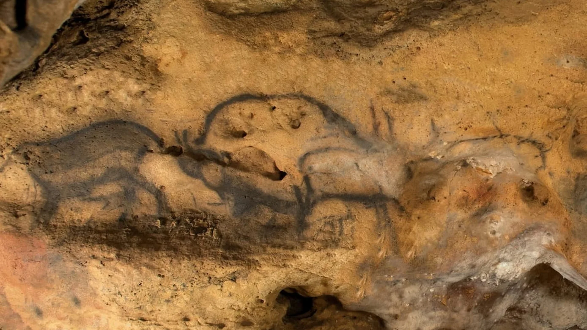
Font-de-Gaume Cave Art Gets Its First Confirmed Radiocarbon Dates
Chemical imaging found charcoal pigment in Font-de-Gaume cave art, letting scientists radiocarbon-date a bison and mask, confirming their Paleolithic age.
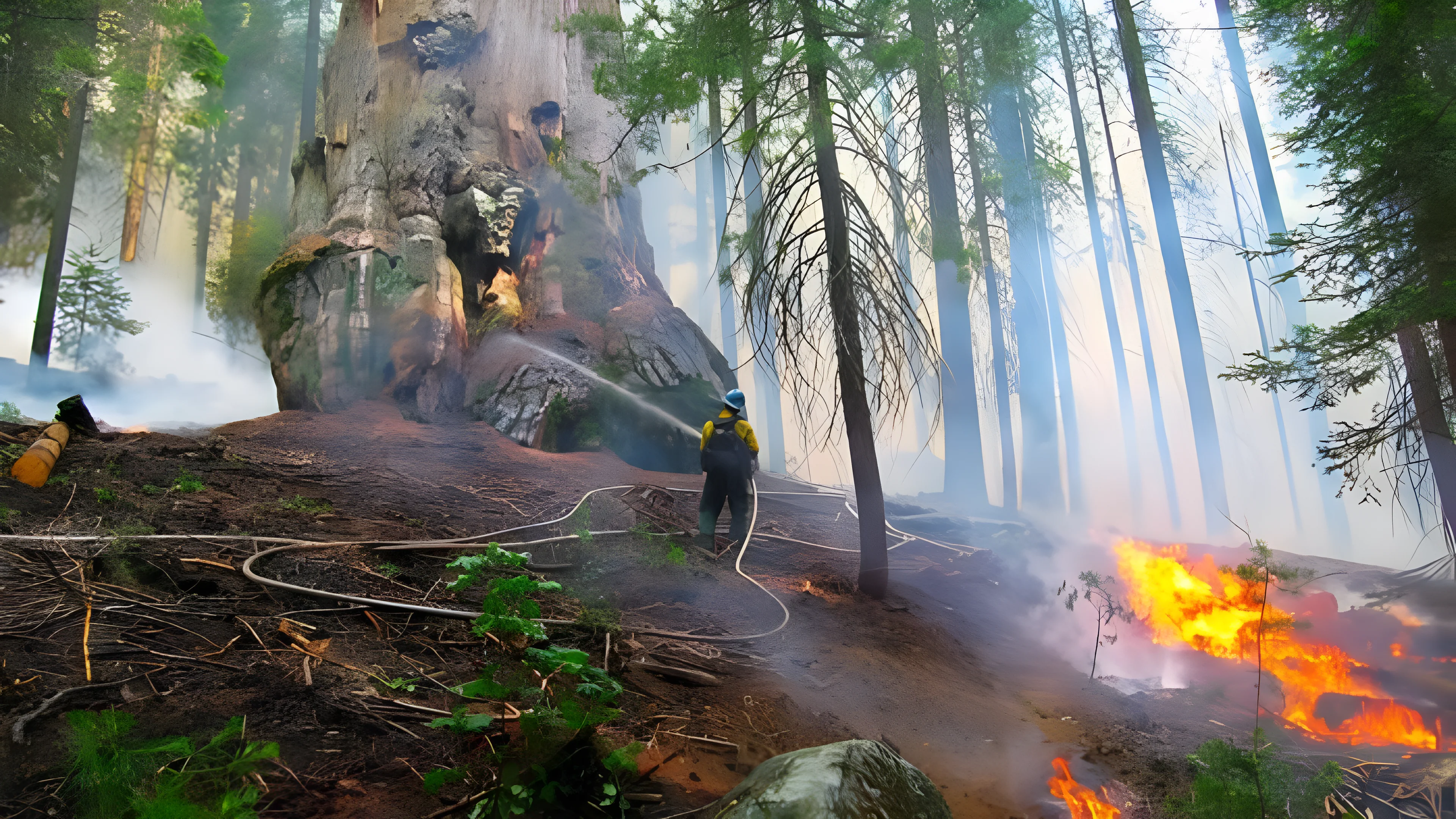
Prescribed Burns Cut Giant Sequoia Wildfire Deaths by 77%, Study Finds
A Nature Communications study of 26,403 giant sequoias found prescribed fire cut wildfire mortality odds 77%, already saving nearly 1,900 ancient trees.
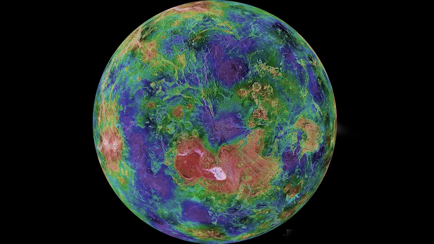
Three Venus Rift Valleys May Be Tectonically Active Today
New 3D models of Venus rifts show extension rates of 3–10 cm/yr; wide flank uplifts at Dali, Ganis and Devana Chasmata suggest active rifting, a new study finds.
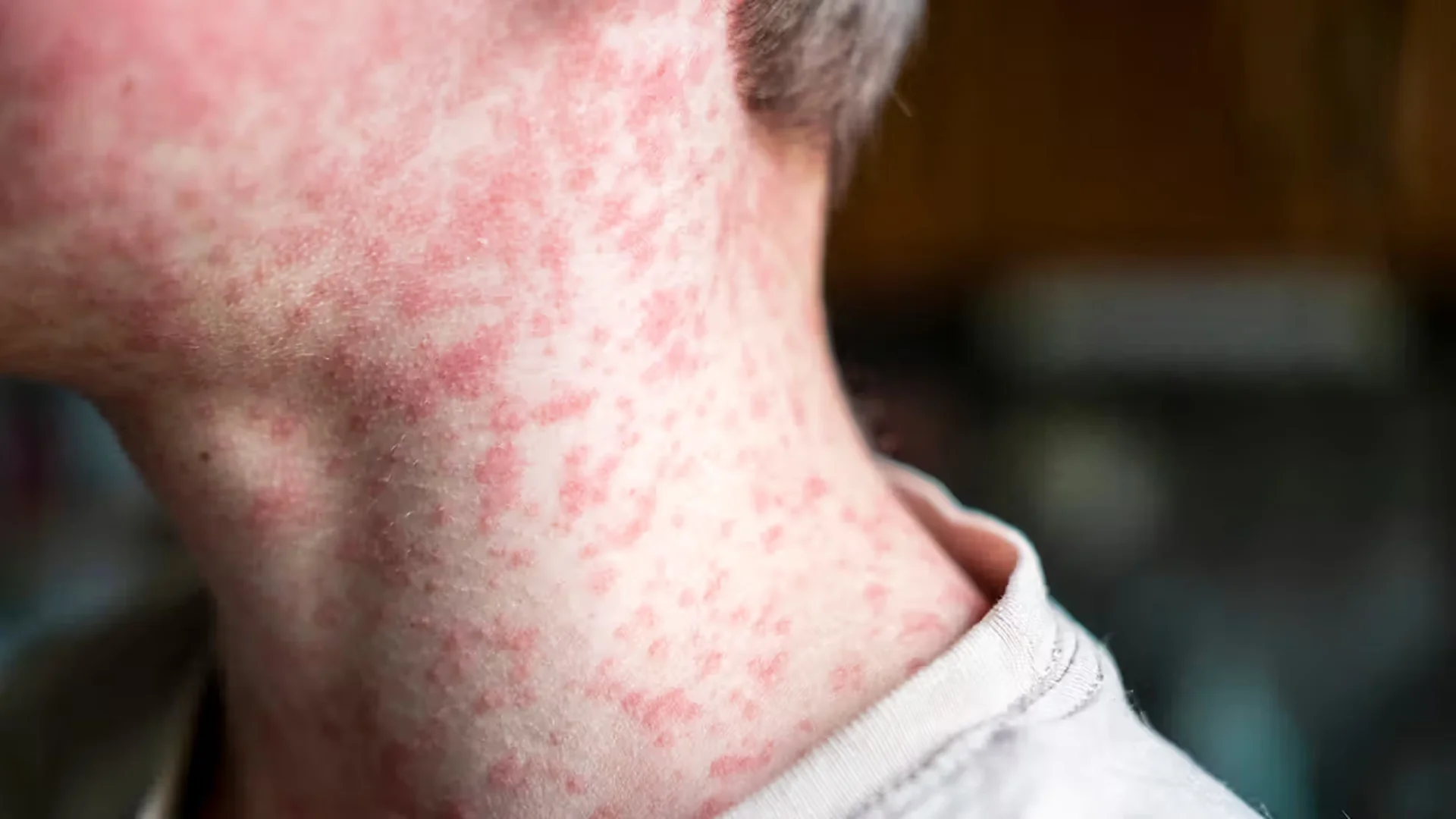
New Antiviral GHP-88310 Blocks Airborne Measles-Like Transmission in Ferret Study
GHP-88310, an oral antiviral, blocked airborne CDV spread in ferrets and cut sources' contagious period by 5 days, a new Nature Microbiology study finds.
Comments (0)
Please sign in to join the discussion.
No comments yet.
Be the first to share your perspective on this topic.