
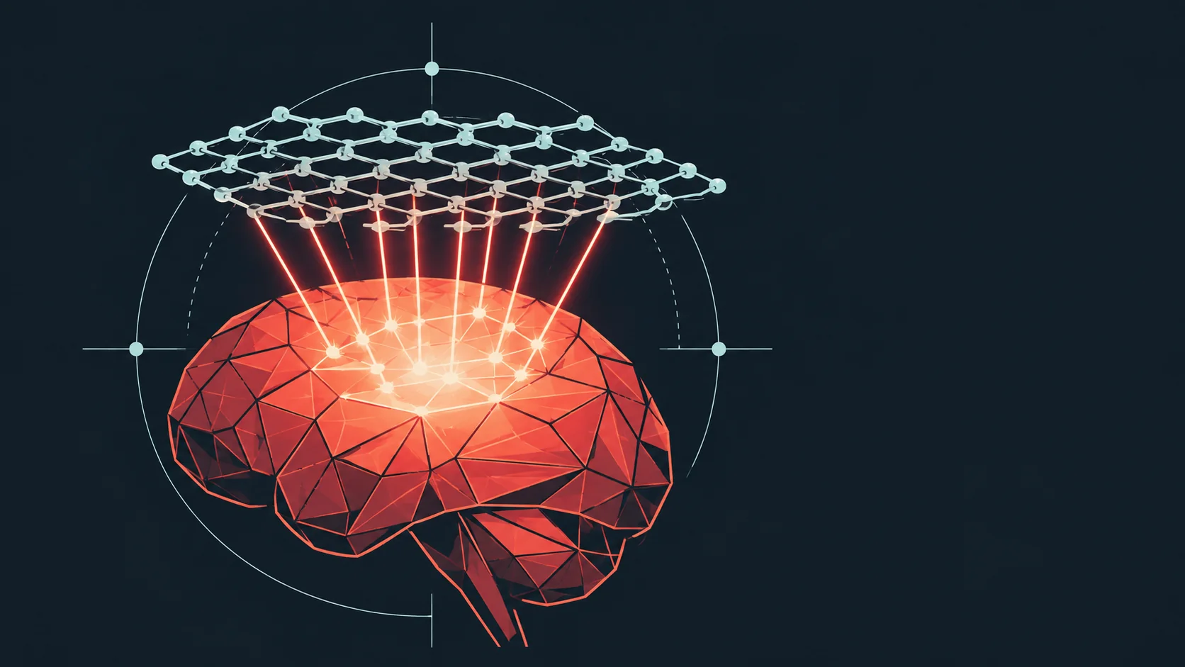
A material engineered from a single-atom-thick sheet of platinum did two separate jobs in sequence during glioblastoma surgery in mice: it lit up tumor clusters too small for current clinical imaging, then, reactivated after the visible tumor was removed, it attacked the cancer cells surgeons could not see or reach.
One Platinum Sheet, Two Jobs, Same Wavelength of Light
Researchers at the University of Technology Sydney, Harvard Medical School, and Henan University built the material as an ultrathin two-dimensional sheet carrying individually placed platinum atoms, positioned using a technique adapted from semiconductor manufacturing. A fluorescent dye attached to the sheet glows under near-infrared light invisible to the naked eye, and a targeting molecule helps the material cross the blood-brain barrier and accumulate specifically in glioma cells. "We've engineered a single material that does two jobs in sequence," said Bingyang Shi, Chair Professor of nanomedicine at UTS's School of Electrical, Mechanical and Biomedical Engineering. "It's a precise guide for the surgeon during the operation, and then a targeted clean-up treatment afterward." During surgery, the material revealed tumor cell clusters as small as 44 micrometers, described by the researchers as a resolution beyond what current clinical imaging tools can achieve. After the visible tumor was removed, the same material was applied inside the surgical cavity and reactivated with the same light. There, platinum atoms convert the tumor's own hydrogen peroxide into oxygen, which counters the low-oxygen environment that normally protects cancer cells from treatment, while the light simultaneously generates heat and reactive molecules aimed at destroying the microscopic cells surgery left behind.
What Surgery Alone Leaves Behind
Glioblastoma's five-year survival rate is roughly 7 percent, and two separate obstacles drive that number: cancer cells spread into surrounding healthy tissue, making complete surgical removal difficult without damaging normal brain function, and the blood-brain barrier limits how well drugs and radiotherapy can reach whatever tumor remains. The mouse results speak to both problems at once, not as separate treatments stacked together but as one material doing sequential work. In the study's glioblastoma mouse model, all mice that received the nanozyme treatment alongside surgery were alive at 60 days. Mice that received surgery alone had a median survival of 42 days. That gap, roughly 18 days beyond the surgery-only baseline within the study's own observation window, is a figure the study reports directly rather than one this article infers, and it lands during exactly the recurrence window that makes glioblastoma so difficult to treat.
Why 60 Days in Mice Isn't a Human Timeline Yet
Shi was direct about the gap between this result and clinical use: "The results are very encouraging, but this is still early-stage research carried out in mouse models, not in people, and that distinction is important. Its imaging and therapeutic performance will also need to be confirmed at the scale of a human brain." A mouse brain is a few centimeters across; light penetration, drug distribution, and surgical navigation all behave differently at human scale, and none of that has been tested yet. The follow-up testing reported so far found no detectable neurological or motor impairment in treated mice, which is a relevant safety signal but not a substitute for toxicology and dosing work in a larger animal or a human trial. If the platform holds up through that process, the researchers frame the eventual goal narrowly: surgeons seeing more of the tumor during the operation, and treating more of what's left behind afterward, rather than a cure for glioblastoma outright.
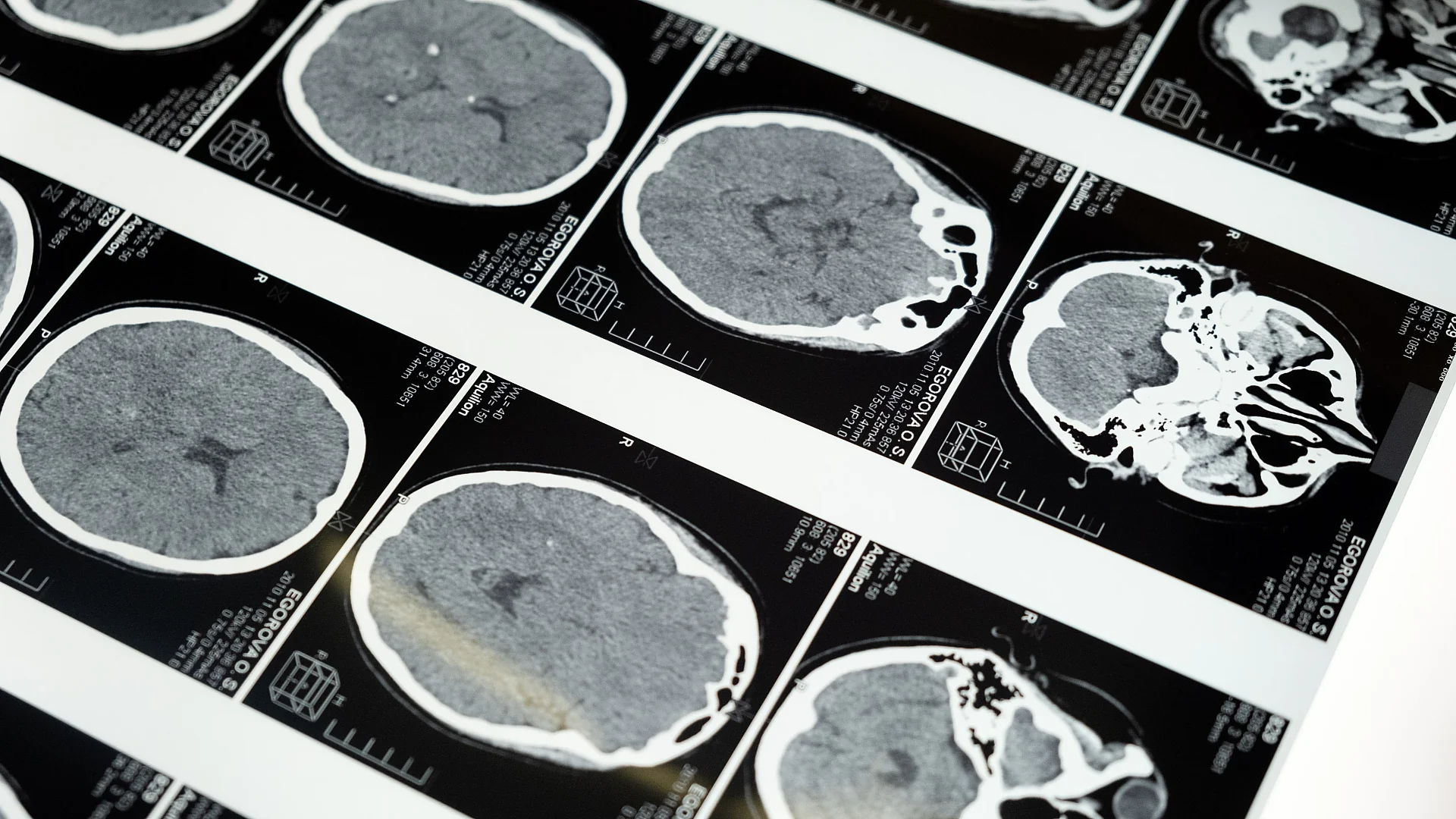
Five Candidate Depression Phenotypes Emerge From MEG Brain Oscillations
MEG depression phenotypes: 263 patients cluster into five oscillation profiles, yet brain–symptom links explain only 3–4% of shared variance in the cohort.

A Seven-Day Water Fast Moved 1,044 Blood Proteins, and Day 3 Was the Turning Point
A 7-day water fast changed 1,044 blood proteins, few before day 3. A second 2025 cohort shared 49 significant hits with no conflicting direction of change.
Paricalcitol and Pancreatic Cancer: Safety Data Published, Phase 2 Not Pursued
Paricalcitol plus gemcitabine and nab-paclitaxel in 36 patients: 42% of the oral arm had grade 2–4 hypercalcemia, and the registry says phase 2 was not pursued.

Mouse and Stem Cell Data Show the Brain Has Two Separate Origins
A Stanford lineage-tracing study finds two separate progenitor cells, marked by Otx2 and Gbx2, build the forebrain/midbrain and hindbrain from gastrulation.
Comments (0)
Please sign in to join the discussion.
No comments yet.
Be the first to share your perspective on this topic.